Introduction:
The modern society we live now value “Appearances of people“ highly due to the evolution of all the markets shifting from conventional physical stores to virtual business model. With rapid growth of social media and platform activities, the presentation of people is no longer through verbal communication or body language but through their photos or videos virtually majority. That brings out the significant importance of smile, which according to the surveys is the top parameter people will be appreciated as there first impression. However, creating an attractive and natural smile is quite challenging to dentists due to some limitation as followings,
(1) Difficulty in transferring or establishing landmarks between working casts and intra/extra oral condition of the patients.
(2) Confusion or misunderstanding of communication among Interdisciplinary team members and laboratory technicians.
(3) Lack of information of patient’s expectation and with limited interaction between patients.
With the assist of modern digital tools and softwares, dentists can superimpose different images and files creating the ideal smile back on patients using the same dental fundamental knowledge but with the actual visual appreciation through simulation on the screen. The digital design could be modified on virtual casts fulfilling parameters of dental esthetics and with the modifying appreciation all together be presented through patients’ clinical photos. With the simulation though all the images, dentists would have much more control in creating the ideal natural smile back for patients avoiding wasting unnecessary clinical time or procedures.
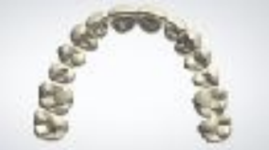
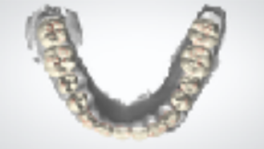
Digital diagnostic wax up based on photos of smile design
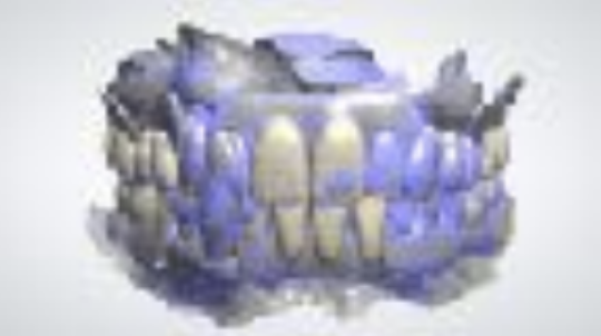
Superimposition of digital diagnostic wax up and intra-oral scan
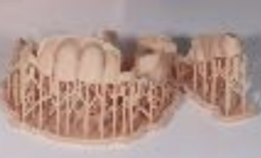
Prototype design based on digital diagnostic wax up
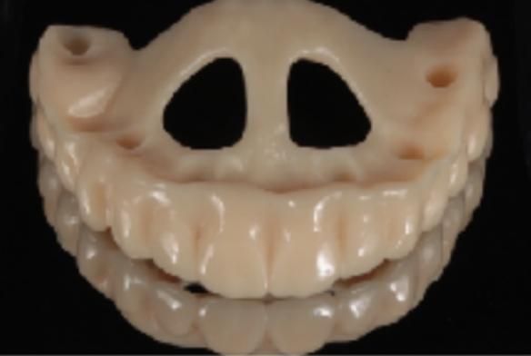
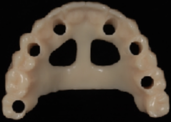
Fabrication of prototype
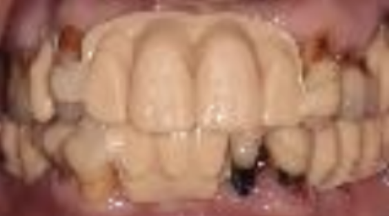
3.PrototypeforClinicalTrial(2ndappointment)
Verifying the fit of prototype
Adjustment of occlusal interference
Verified facial profile
Mark the adjustments and re-scan
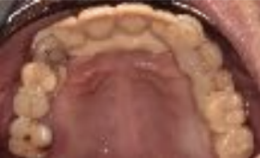
4.FabricationofProvisional/DefinitiveProsthesis
ClinicalProcedures: Step1/DataCollection:
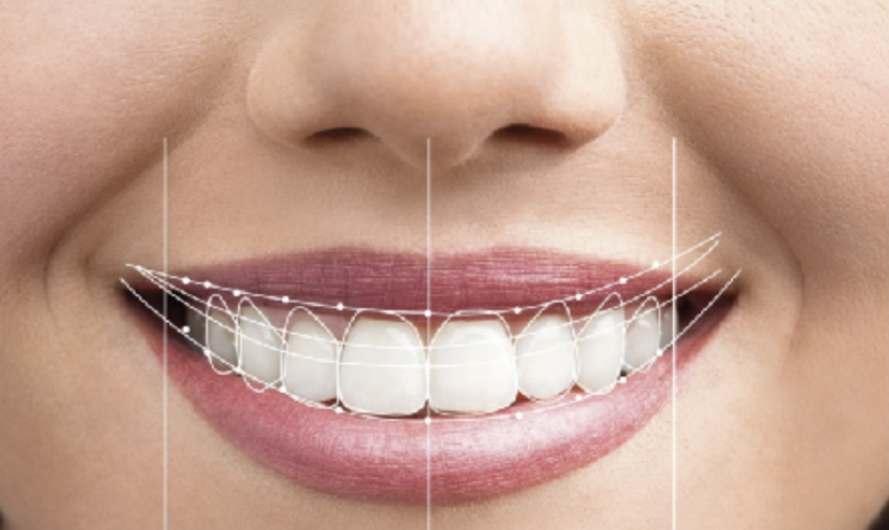
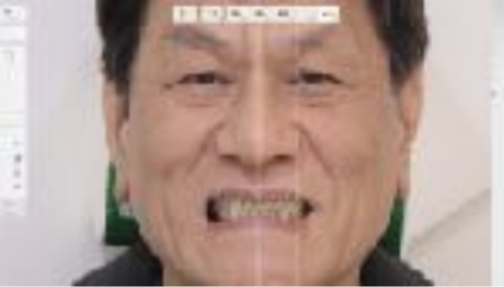
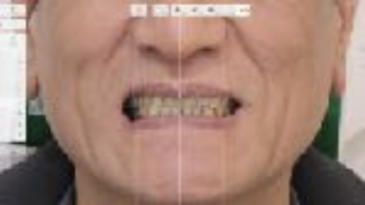
Design ideal anterior teeth position, proportion and alignment using smile design software (3Shape Smile Design®). Clinical photos with specific shooting were taken following by the recommendation of the manufacturer.
Esthetic parameters for establishing ideal smile were as followings,
1. Determine incised edge position
The exposure of teeth at rest position (pronounce “Emma”)
Phonetics (“V”/ “S” sound)
2. Determine midline, distal line of canine
3. Determine lip suport
4. Determine plane of occlusion (parallel to inter-pupillary line; curvature of smile)
5. Define the free gingival margin (Zenith) using ideal proportion of anterior teeth
6. Virtual wax up anterior teeth by ideal proportion and axis
7. Established reasonable buccal corridor
Step2/ComputerAidDesign:
Diagnostic wax up was done by using 3Shape Dental System® following by the
esthetic parameters discussed through step 1.
1. Previous dental prosthesis can be a reference guide also assisting the digital design re-establishing new landmarks.
2. By setting the previous prosthesis as “pre-scan” in the designing software, the pre- scan template act as a physical guide (in color blue of design) allowing us verifying the ideal teeth position with ease while processing digital waxing up.
3. At the end, we could have a full smile view simulating digital wax up transferring
back to patients.
Step 3/ Prototype for Clinical Trial:
1. Prototype from digital design was printed out using 3D printer (Formlab2).
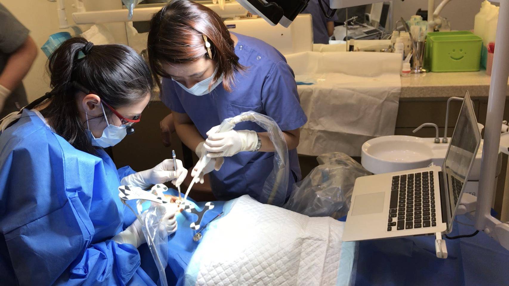
2. Clinical try-in was carried out for verifying the fit of maxillary and mandibular prototypes following by checking the anterior teeth position, lip support and phonetics.
3. Plane of occlusion, treatment position and occlusal scheme can also be adjusted on the prototype through the trial sessions providing solid landmarks and real visual appreciation for both dentists and patents.
Step 4/ Fabrication of provisional or definitive prosthesis:
Conclusion:
With the aid of modern digital equipments and softwares, the digital smile design can be much more relatable to the clinical scenario of ideal smile. By following the digital workflow, smile prototypes, provisional restorations, and definitive prosthesis could be planned and fabricated based upon the same digital diagnostic wax up. Predictable and accurate outcome can be achieved with the protocol which makes diagnosis more efficient and treatment plans more consistent. It will also provide more logical and straightforward treatment sequences reducing the risks and improving the natural results.
References:
1.Ward DH. Proportional smile design using the RED proportion. Dent Clin North Am 2001;45(1):143-154.
2.Ward DH. Using the RED Proportion to Engineer the Perfect Smile. Dentistry Today 2008:48-53
3.Chu SJ. A biometric approach to predicatable treatment of clinical crown discrepancies. Pract Proced Aesthet Dent 2007;19(7):401-408
4.Chu SJ, Tan JH, Stappert CF, Tarnow DP. Gingival Zenith Positions and Levels of the Maxillary Anterior Dentition. J Compilation 2009; 21(2):113-120
5.Liu X, Yu J, Zhou J, Tan J. A digitally guided dual technique for both gingival and bone resection during crown lengthening surgery. J Prosthet Dent 2018;119(2):345-349 6.Scutella F, Landi L, Stellino G, Morgano S. Surgical template for crown lengthening: A clinical report. J Prosthet Dent 1999; 82(3):253-256
7.Machado A, Mccomb R, Moon W, Gandini L Jr. Influence of the Vertical Position of Maxillary Central Incisors on the Perception of Smile Esthetics Among Orthodontists and Laypersons. J Esthet Restor Dent 2013; 25(6):392-40
8.Spear FM, Kokich VG, Mathews DP. Interdisciplinary management of anterior dental
esthetics. J Am Dent Assoc 2006;137(2):160–9.
9.Chu SJ. Range and mean distribution frequency of individual tooth width of the maxillary anterior dentition. Pract Proced Aesthet Dent 2007;19(4):209–15.


.png)